






Breakthrough in Microscopy Technology Announced by Physicists at University of California, Berkeley
Physicists at the University of California, Berkeley, have developed a device called a laser phase plate that uses a similar principle as phase contrast microscopy to improve cryoelectron microscopy.
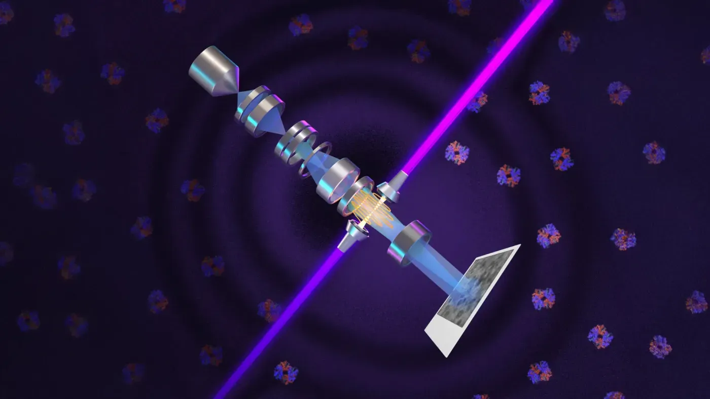
A breakthrough in microscopy technology has been announced by physicists at the University of California, Berkeley. They have developed a device called a laser phase plate that uses a similar principle as phase contrast microscopy, which was introduced nearly a century ago.
This technique made it possible to study cellular structures more easily and earned a Nobel Prize in 1953. The researchers' new innovation brings this same principle to electron microscopy, allowing for the magnification of objects up to 10,000 times more than conventional light microscopes can achieve.
The device is designed to improve cryoelectron microscopy (cryo-EM), which has become a crucial tool for studying protein structures and speeding up drug discovery over the past decade. However, it still faces significant challenges due to its limitations in imaging small molecules.
The new laser technology could overcome this obstacle by allowing scientists to image proteins as small as one-third their current limit. This would be a major breakthrough in understanding how these complex molecules function within living cells.
Cryoelectron tomography (cryo-ET) is another technique that combines images from multiple angles to create three-dimensional views of proteins and other molecules. Unlike techniques that study isolated molecules, cryo-ET can reveal the arrangement and interaction of proteins within their natural cellular surroundings.
The development of this laser phase plate has the potential to significantly improve the resolution of both cryo-EM and cryo-ET methods. This could lead to a greater understanding of how biological macromolecules function together in living cells.
Physicists at UC Berkeley are optimistic about the impact of this new technology, which they believe will overcome key limitations in imaging small molecules. The researchers expect their laser phase plate to increase signal-to-noise ratio and allow for the analysis of previously inaccessible proteins.
The researchers have designed a new microscope that could significantly improve image quality and contrast. This breakthrough relies on a highly intense focused continuous-wave laser, which interacts directly with the electron beam to alter its phase.
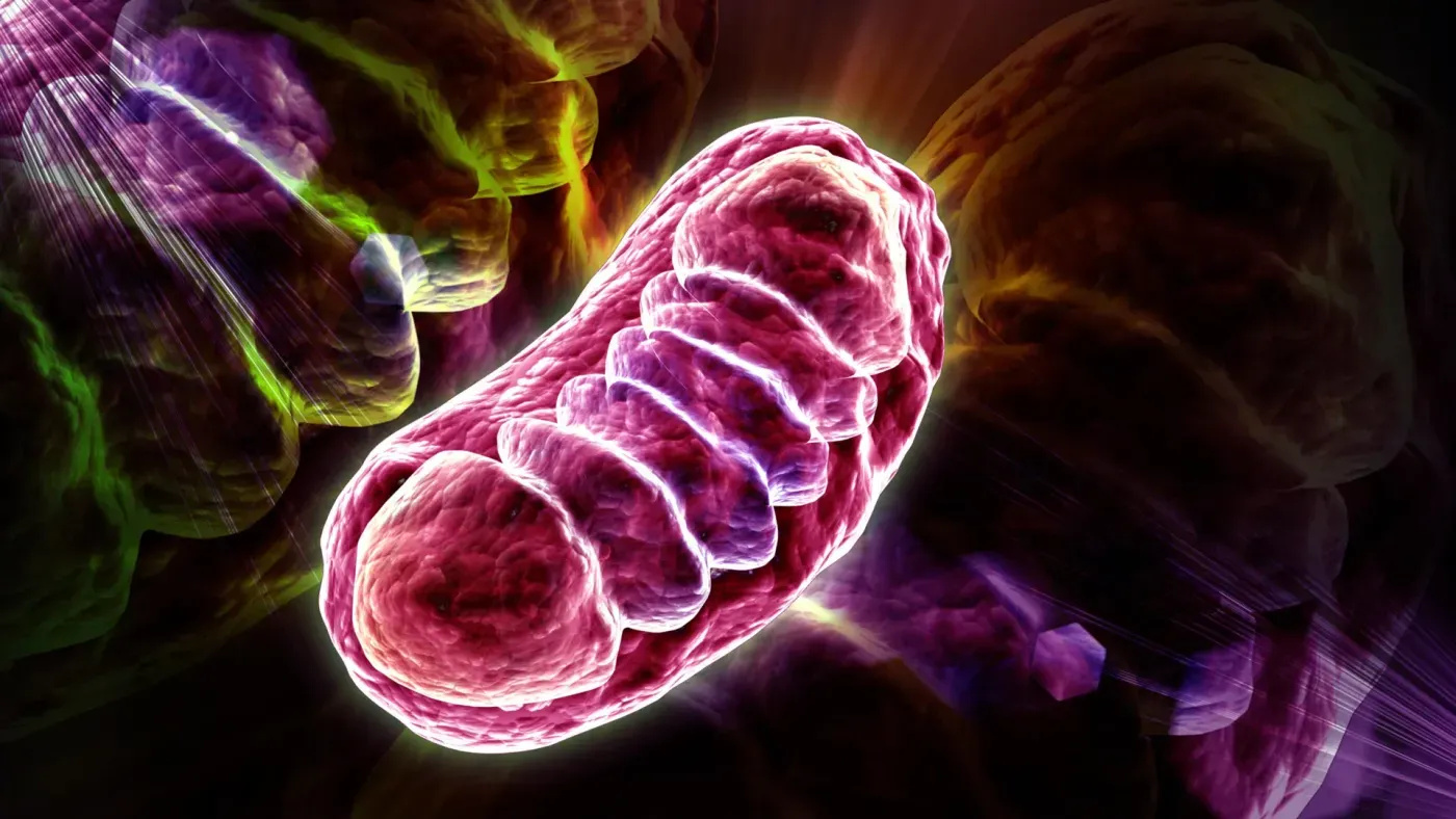
This adjustment enhances the ability to distinguish small molecules from their surroundings, including hemoglobin, and improves visibility of structures inside cells like the nucleus and mitochondria. The improved contrast is essential for studying complex cellular material.
Cryogenic electron tomography (cryo-ET) requires a significant increase in image contrast to visualize intricate cellular components. Bridget Carragher, founding technical director of imaging at Biohub, notes that cryo-ET involves examining crowded cellular material, akin to searching for a single leaf on a tree within a dense forest.
The new microscope features an advanced cryo-EM instrument equipped with the laser phase plate. This setup is expected to provide a dramatic improvement in contrast, enabling researchers to visualize previously inaccessible structures and molecules inside cells.
Müller has named his microscope "Theia", after the ancient Greek Titaness associated with light and radiance. Theia is an advanced cryo-EM instrument that offers enhanced resolution capabilities even without the laser phase plate.
Meanwhile, Bridget Carragher's team at Biohub is constructing a related microscope using two lasers instead of one. This design builds on theoretical research conducted by Müller and his collaborators.
The two-laser setup involves arranging the beams at right angles to each other, with each operating at approximately half the power of Theia. This configuration helps prevent component burnout and reduces optical distortions that could compromise image quality.
The limitations of current microscopy technology have long been a challenge for scientists studying cellular structures. One major hurdle arises from the transparency of plant and animal cells when viewed under normal light conditions. This is because these cells are comprised mostly of water, which scatters very little light and makes it difficult to discern small features.
To overcome this issue, researchers have traditionally resorted to staining techniques that add contrast but unfortunately often come at a cost: cell death. This has led scientists to seek alternative approaches for creating clear images of cellular structures without harming the cells themselves.
A major breakthrough in microscopy was achieved by Dutch physicist Frits Zernike in 1930. He discovered that light passing through biological material undergoes changes beyond mere brightness or dimness, including a phase shift caused by scattering within the cell. This tiny delay alters the timing of the light wave peaks.
Zernike's realization led to the development of phase-contrast microscopy, where the phase of unscattered light is shifted by 90 degrees to enhance contrast and reveal subtle cellular features. His innovative technique earned him the Nobel Prize in Physics in 1953, a testament to its significance for biological research.
The potential of phase-contrast microscopy was quickly recognized by scientists in the early 1940s, who began exploring ways to adapt this technology for electron microscopes. These devices use electrons instead of light to examine structures as small as individual proteins, offering unparalleled resolution and detail.
However, initial attempts to apply phase contrast to electron microscopes were hindered by technical difficulties. Devices designed to alter the phase of electron beams often compromised beam strength or introduced image instability, limiting their effectiveness in producing clear images.
Müller's and Glaeser's innovative approach to phase shift used an intense laser to produce the necessary phase change without compromising electron beam intensity.
This solution offered a significant improvement over previous methods that often compromised beam strength or introduced image instability, limiting their effectiveness in producing clear images.
Glaeser has made substantial contributions to the development of cryo-EM, which presents a potentially simpler way to determine molecular structures than traditional X-ray crystallography.
Cryo-EM avoids the need to crystallize molecules, but it poses its own set of challenges, including difficulties with specimen preservation during imaging.
The biggest problem in cryo-EM is that an electron beam can heat and damage the specimen being examined, limiting the amount of information that can be collected and the level of detail in the resulting images.
To mitigate this issue, researchers have explored various methods, including covering specimens with metal to reduce damage and improve contrast. However, this approach also reduces image sharpness.
During the 1960s, scientists began investigating whether freezing biological samples could protect them from radiation damage during imaging.
Glaeser's research demonstrated that cooling specimens helps preserve them during imaging, and he proposed using a less intense electron beam to examine thousands of frozen molecules at once, further reducing damage.
Computers can analyze and combine the images of multiple molecules positioned at different angles to reconstruct a highly detailed molecular structure. This approach has been instrumental in advancing the field of modern cryo-EM.
The developers of cryo-EM were recognized with the 2017 Nobel Prize in Chemistry for their groundbreaking work, which was acknowledged by the Nobel laureates during their acceptance remarks as a significant advancement in biochemistry.
The development of a revolutionary new microscope has been ongoing for over a decade, driven by the need for an incredibly intense laser beam focused into a tiny area.
Generating such a concentrated beam was the first major hurdle that researchers needed to overcome in order to change the phase of an electron beam by 90 degrees.
To achieve this feat, the team employed a novel approach: confining a continuous laser beam within a spherical chamber lined with mirrors. This clever design allowed the light to repeatedly reflect through the cavity more than 10,000 times, significantly increasing its intensity while focusing it into an extremely small spot.
The resulting beam is an astonishing 75 kilowatts of power focused onto a few microns, equivalent to the energy output of a welding laser or even a military-grade laser. This monumental achievement has led to the creation of the brightest continuous laser focus ever recorded.
The team first demonstrated the effectiveness of this concept by integrating a laser phase plate into an older electron microscope belonging to Glaeser.
Subsequent funding from Biohub enabled them to acquire a state-of-the-art Thermo Fisher Krios cryo-EM microscope, which they then modified to accommodate the innovative laser technology.
In their latest research, the scientists put this upgraded microscope through its paces using six diverse samples with varying sizes and preparation methods. The results showed that the laser consistently improved image resolution, particularly for challenging specimens like small particles or poorly prepared samples.
The introduction of a laser phase plate has significantly improved the imaging capabilities of cryo-EM microscopes.
Proteins are complex molecules that play crucial roles in various cellular processes, but their structures can be difficult to visualize due to their small size and poor sample quality. Researchers have long been searching for ways to overcome these challenges, particularly when dealing with proteins below 70 kilodaltons in mass. This is a critical limitation, as such proteins account for approximately 90% of the human proteome.
The new laser phase plate technology has expanded the capabilities of cryo-EM microscopes, enabling researchers to image proteins as small as 50 kilodaltons. While imaging these tiny proteins remains a challenging task, this breakthrough has opened up new avenues for scientific inquiry. Proteins below 70 kilodaltons are particularly significant, as they make up the vast majority of the human proteome.
The potential implications of this technology are far-reaching, with researchers anticipating that it could eventually allow scientists to study even smaller proteins, such as myoglobin, which has a mass of around 17 kilodaltons.
Facts based on reporting originally published by ScienceDaily Health.
You may republish this story, in full or in part, if you credit Noti Group and link to it (licence CC BY 4.0). Photos are not included.